Specialised Pelvic Ultrasound for Women
(Gynaecological, Fertility, and Pregnancy)

Looking for real answers and solutions?

We work with Perth’s leading specialist women’s sonographer with the most sensitive and accurate technology in Western Australia to provide insight into many areas of female gynaecological conditions, anatomy, and female fertility structures.
These are areas standard women’s pelvic ultrasound providers are not trained in; and are not able to accurately view, measure, or assess with standard pelvic ultrasound equipment.

For just one example, we work with the very first ultrasound practice in WA to establish sonovaginography for the detailed assessment of severe endometriosis via ultrasound (this is usually or historically only able to be assessed upon a surgical laparoscopy), among many other conditions, structures, and concerns which are not provided or able to be detected on standard pelvic ultrasounds.

Specialist and detailed pelvic ultrasound services including:

  • Specialist pelvic ultrasound through WA’s leading specialist women’s health sonographer; a female registered Obstetrics & Gynaecology Ultrasound Specialist and Consultant Sonologist.
  • Accurate and sensitive measurement of the endometrium thickness, which plays a role in both fertility and the assessment of perimenopause/menopause.
  • The number of follicles and developing follicles within each ovary, including their measurements of size, which are not commonly able to be viewed upon standard pelvic ultrasound equipment.
  • This sensitive assessment may be utilised for fertility patients, as well as assessing the degree of perimenopause or menopause stage within each ovary which many of our women patients may utilise to assist their decision to halt the OCP or have their Mirena IUD removed if they are unsure if they have finished menstruating or have reached menopause.
  • Confirmation as to whether ovulation is occurring; and confirmation as to whether ovulation is occurring from the left or right ovary that month. This is especially important in certain fertility patients who only have one fallopian tube or have a partial tubal blockage such as a hydrosalpinx present.
  • Examination of fallopian tubes for health and any possible blockage/s such as a hydrosalpinx being present (fallopian tube blockage due to fluid build-up) or blockage due to a past pelvic infection (often previously undiagnosed) and allow us to refer for appropriate gynaecological intervention if this is required.
  • Assessment of hyperechoic areas present within each ovary, any areas of calcifications within the ovaries, acoustic shadowing present within the ovaries, and the presence of any follicular cysts.


We are proud to refer our patients to the highest quality service of women’s specialist pelvic sonography (ultrasound) through the only WA leading provider with state-of-the-art equipment including high end imaging capabilities using 2D and 3D/4D ultrasound technology offering comprehensive ultrasound services in both Obstetrics (pregnancy ultrasounds) and Gynaecology (non-pregnancy pelvic ultrasounds).

Detection and measurement of female pelvic organs and health:

pexels-victoria-borodinova-1648374
  • Sensitive 3-dimensional measurement and volume of the size of both bilateral ovaries.
  • Accurate and sensitive assessment of the size, position, and contour of the uterus.
  • Precisely identify adenomyosis within the uterus muscle walls, which if present may cause symptoms of abnormal or heavy menstrual bleeding painful periods, pain with intercourse, (dyspareunia), bleeding between periods, and increased iron loss.
  • Assessment of endometrium-myometrial junction, which is not provided on standard pelvic ultrasounds.
  • Detection of pelvic congestion syndrome, which in simple terms means varicose veins within the pelvic cavity, including dilated arcuate veins and both left and right adnexal veins, and the specific measurement of these dilated or varicose veins to their maximum millimetre measurement (i.e., measuring up to 9.9mm), which is not provided on standard pelvic ultrasounds. Pelvic congestion syndrome is an often-missed diagnosis in Australia as it is not able to be viewed or measured upon on standard pelvic ultrasound equipment. Pelvic congestion syndrome is commonly associated with period pain (dysmenorrhea), pelvic pain, heavier periods (menorrhagia), a heaviness within the pelvic areas or legs, and increased iron loss.
  • Pain and tenderness scores for each pelvic cavity organ, including cervix, uterus, both ovaries, bilateral uterosacral ligaments, Pouch of Douglas, bilateral adnexal veins, adnexal region, utero-vesical fold, and a small area of the lower bowel, which are not provided on standard pelvic ultrasounds, and may indicate endometriosis and/or the presence of adhesions or tethering to other pelvic cavity organs such as the bowl.
  • Comprehensive mobility assessment of each pelvic cavity organ, including uterus, both ovaries, bilateral uterosacral ligaments, Pouch of Douglas, bilateral adnexal veins, utero-vesical fold, and a small area of the lower bowel, which are not commonly provided on standard pelvic ultrasounds, and may indicate endometriosis or adhesions tethering to these organs.
  • Evidence of thickening and/or nodules of both uterosacral ligaments, which is not provided on standard pelvic ultrasounds.
  • Accurate and sensitive assessment for nabothian cysts on the cervix, including the measurement of size of nabothian cysts and number present, which is not provided on standard pelvic ultrasounds.
  • Including assessment of endocervical polyps or feeder vessel/s, which is not provided on standard pelvic ultrasounds.
  • Accurate and sensitive assessment for evidence of endometriomas or ovarian cyst/s, which are not able to be accurately assessed on standard pelvic ultrasounds.


Our practice is proud to refer for specialist pelvic ultrasound through WA’s leading specialist women’s health sonographer who is a female registered Obstetrics & Gynaecology Ultrasound Specialist and Consultant Sonologist committed to the highest levels of care, alongside her team of experienced, kind, and genuinely caring support staff.

Accurate assessment & measurement of fibroids, cysts, polyps, & more:

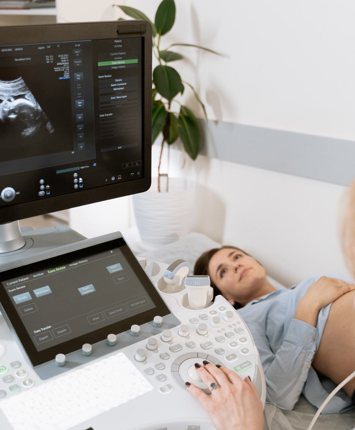
We provide precise and comprehensive assessment of the presence and measurement of uterine fibroids, cervical cysts, and endometrial polyps which are not commonly able to be viewed upon standard pelvic ultrasound equipment.

The includes the in-depth measurement with spectral doppler for peripheral and internal vascularity of each endocervical polyp/s and uterine fibroid/s; the resistive index of each polyp/s and fibroid/s; and the peak systolic velocity of each polyp/s and fibroid/s.

These may also play a role in fertility and gynaecological symptoms such as period pain, heavier periods, pain upon intercourse, miscarriage, and increased iron loss, which are not accurately provided on standard pelvic ultrasounds, including:

pexels-andre-furtado-1263986
  • Intramural fibroids
    • Intramural fibroids appear in the muscular wall of the uterus and are the most common type of fibroid. Due to the location of intramural fibroids, they may grow larger and can stretch the womb.
  • Subserosal fibroids
    • This type of fibroid grows outside of your uterus on the serous membrane (or serosa), which is the outer lining that all organs and internal body cavities have. Subserosal fibroids may grow big enough that they can make your womb look bigger on one side.
  • Pedunculated fibroids
    • Pedunculated fibroids form when a Subserosal fibroid develops a stem. This stem turns into a slender base that can support a tumour. If this happens, this is where a pedunculated fibroid grows.
  • Submucosal fibroids
    • Submucosal fibroids grow in the myometrium, or middle layer of muscle in the uterus. This type of fibroids is not as common as the other three.


A referral to our specialist women’s pelvic ultrasound may be organised partially through Medicare, or privately referred through our practice.

We are here to support your fertility and gynaecologist health, working alongside your medical GP and gynaecologist specialist or referring you to one of our recommended doctors and specialist gynaecologists to fit your individual needs. Combining sophisticated health testing solutions with the traditional wisdom and power of natural medicine alongside our Australian medical system!

Let us guide you to a happier, healthier you.